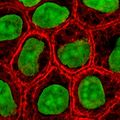
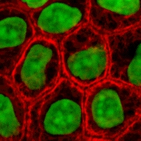
ဖိုင်:Epithelial-cells.jpg
Epithelial-cells.jpg (၂၀၂ × ၂၀၂ pixels, ဖိုင်အရွယ်အစား - ၄၆ KB, MIME အမျိုးအစား image/jpeg)
ဖိုင်မှတ်တမ်း
ဖိုင်ကို ယင်းနေ့စွဲ အတိုင်း မြင်နိုင်ရန် နေ့စွဲ/အချိန် တစ်ခုခုပေါ်တွင် ကလစ်နှိပ်ပါ။
| နေ့စွဲ/အချိန် | နမူနာပုံငယ် | မှတ်တမ်း ဒိုင်မန်းရှင်းများ | အသုံးပြုသူ | မှတ်ချက် | |
|---|---|---|---|---|---|
| ကာလပေါ် | ၁၉:၄၉၊ ၂ မေ ၂၀၀၅ |  | ၂၀၂ × ၂၀၂ (၄၆ KB) | Helix84 | Cultured MDCK epithelial cells were stained for keratin, desmoplakin, and DNA. The stained cells were visualized by scanning laser confocal microscopy. The image shows how keratin [[Cytoskeleton|cytoskele |
ဖိုင်သုံးစွဲမှု
အောက်ပါ စာမျက်နှာ သည် ဤဖိုင်ကို အသုံးပြုထားသည်:
ဂလိုဘယ် ဖိုင်သုံးစွဲမှု
အောက်ပါ အခြားဝီကီများတွင် ဤဖိုင်ကို အသုံးပြုထားသည်-
- af.wikipedia.org တွင် အသုံးပြုမှု
- anp.wikipedia.org တွင် အသုံးပြုမှု
- ar.wikipedia.org တွင် အသုံးပြုမှု
- az.wikipedia.org တွင် အသုံးပြုမှု
- bg.wikipedia.org တွင် အသုံးပြုမှု
- blk.wikipedia.org တွင် အသုံးပြုမှု
- br.wikipedia.org တွင် အသုံးပြုမှု
- bs.wikipedia.org တွင် အသုံးပြုမှု
- ca.wikipedia.org တွင် အသုံးပြုမှု
- ca.wikibooks.org တွင် အသုံးပြုမှု
- cs.wikipedia.org တွင် အသုံးပြုမှု
- cv.wikipedia.org တွင် အသုံးပြုမှု
- da.wikipedia.org တွင် အသုံးပြုမှု
- el.wikipedia.org တွင် အသုံးပြုမှု
- en.wikipedia.org တွင် အသုံးပြုမှု
- User:JWSchmidt
- Tissue engineering
- Intermediate filament
- User:JWSchmidt/Images
- Wikipedia:Facebook directory
- Cytokeratin
- User:Lexor/Temp/Cell (biology)
- Cell culture
- Metabolic engineering
- User:ClockworkSoul/userpage/panel center
- User:ClockworkSoul
- User:Allthesestars/panel centre
- User:SnapJag/userpage/panel center
- Wikipedia talk:WikiProject Molecular Biology/Molecular and Cell Biology/Discussion Archive
- User:Julie Zamostny/BIO134: Cancer Biology
- User:Mesoderm/sandbox
- User:S spoerri/sandbox
- User:Randoperson1/userboxes
- en.wikibooks.org တွင် အသုံးပြုမှု
- en.wiktionary.org တွင် အသုံးပြုမှု
- es.wikipedia.org တွင် အသုံးပြုမှု
ဤဖိုင်ကို အခြားနေရာများတွင် အသုံးပြုထားမှုများအား ကြည့်ရှုရန်။

